
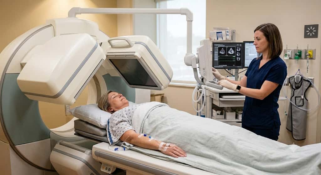
Everything you need to know about nuclear cardiology stress testing, from preparation and imaging to results, PET vs. SPECT, and common questions.
If your doctor just told you that you need a nuclear stress test, you probably have two reactions at once: relief that someone is finally taking a closer look at your heart, and a little unease at the word nuclear. That's fair. Nobody hands you a pamphlet that actually answers the questions you're thinking: How much radiation? What does the injection feel like? Why did your neighbor get a different version of the test than you did?
What Is Nuclear Cardiology Stress Testing?
Nuclear cardiology stress testing, also called myocardial perfusion imaging (MPI), measures blood flow through the heart muscle both at rest and during exertion. A small amount of radioactive tracer is injected into your bloodstream, and a special camera picks up the signal it gives off as it moves through your heart. Source: MedlinePlus
Areas getting good blood flow light up clearly. Areas with reduced flow, usually because a coronary artery is narrowed or blocked, show up dimmer. This is different from a standard treadmill stress test, which only tracks your heart's electrical activity (EKG) and symptoms while you exercise.
A regular stress test can miss blockages that a resting EKG looks normal for. Adding the nuclear imaging component lets your cardiologist actually see the blood flow itself, which is why it's often the next step when a basic stress test is inconclusive or when your symptoms and risk factors warrant a closer look from the start.
Why Do Doctors Order This Test?
The most common reasons this test gets ordered:
Chest pain or shortness of breath that could be caused by reduced blood flow to the heart
Known coronary artery disease, to see how much it's affecting blood flow now
After a heart attack, to check how much heart muscle is still getting adequate blood supply
Before major surgery, to clear you from a cardiac standpoint if you have risk factors
Following up on an abnormal or ambiguous EKG stress test
Here's the part that often gets left out: this isn't automatically the right first test for everyone with chest pain. Current cardiology guidelines emphasize matching the test to the patient.
Someone with a low probability of coronary artery disease might be better served by a coronary CT angiogram (CCTA), which looks directly at the arteries rather than blood flow. Someone who can't tolerate radiotracers or has kidney issues might get a stress echocardiogram instead. Source: ASNC
SPECT vs. PET Nuclear Stress Tests: What's the Difference?
There are two main technologies, and they're not interchangeable; they use different equipment and tracers and have different strengths.
Single-Photon Emission Computed Tomography (SPECT)
SPECT (Single-Photon Emission Computed Tomography) is the more widely available option. It typically uses technetium-99m-based tracers (brand names include Cardiolite and Myoview). Most hospitals and imaging centers have SPECT cameras, so this is what most people get.
Positron Emission Tomography (PET)
PET (Positron Emission Tomography) uses a different tracer, most commonly rubidium-82, and produces sharper images faster. PET is particularly useful for patients with larger body size, for whom SPECT images can be harder to interpret, and it generally involves less radiation exposure per scan. The tradeoff is availability: fewer centers have cardiac PET capability, and it tends to cost more.
If you're a candidate for either, your cardiologist's choice usually comes down to your body type, local equipment availability, and how quickly you need results. Neither is better across the board; they're suited to different situations.
Exercise vs. Pharmacologic Stress: How the Stress Part Works?
Stress just means making your heart work harder than it does at rest, and there are two ways to do that.
Exercise stress
Exercise stress means walking on a treadmill with an increasing incline and speed, similar to a standard stress test, while the tracer is injected near peak exertion.
Pharmacologic stress
Pharmacologic stress uses medication instead of exercise, for patients who can't walk on a treadmill due to joint problems, poor conditioning, or other limitations. The most commonly used drugs are:
Regadenoson: A vasodilator that widens coronary arteries in a way that mimics the effect of exercise. It's the most commonly used agent today because it's given as a quick injection with a short duration of action.
Adenosine: An older vasodilator with a similar mechanism, given as a continuous infusion. It's used less often now that regadenoson is available, partly because its effects can cause more noticeable chest tightness or flushing during the infusion.
Dobutamine: It's used when vasodilators aren't a good option, often because of significant asthma or COPD. It works by directly increasing heart rate and contraction strength rather than dilating arteries.
Knowing which one you're getting matters, because the side effects differ. Vasodilators can cause a brief flushed, warm feeling or mild shortness of breath. Dobutamine can cause a racing heartbeat or jitteriness, similar to a caffeine rush. Both wear off within minutes of stopping the medication. Source: Cleveland Clinic
How to Prepare for Your Nuclear Stress Test?
The standard instructions are familiar: no caffeine, no food for several hours, hold certain medications. But why do these rules actually exist? Skipping them isn't just ideal; it can invalidate the test.
Caffeine and theophylline-containing drugs: Block the same receptors that regadenoson and adenosine work on. If caffeine is still in your system, the vasodilator won't work properly, and the test may need to be rescheduled. This includes decaf coffee, which still has small amounts of caffeine, and some pain relievers and sodas that contain it too.
Beta-blockers: Slow your heart rate, which is exactly what they're prescribed for, but that also blunts your heart's ability to reach the target rate needed for a valid exercise test. Most cardiologists will have you hold these for 24–48 hours beforehand, but never stop a prescribed medication without your doctor's specific instruction to do so.
Sildenafil and similar erectile dysfunction medications: They cause a dangerous drop in blood pressure when combined with nitrates sometimes given during the test, so these need to be disclosed and typically held for 24- 48 hours.
What Happens During the Test?
Plan for 3 to 5 hours total, even though the active parts of the test are much shorter. Here's a realistic walkthrough:
IV placement and a resting tracer injection, followed by a wait of 30–60 minutes for it to circulate before the first set of images.
Resting images are taken, which takes about 15–20 minutes lying still under the camera.
Stress phase: either treadmill walking with increasing intensity or a pharmacologic infusion lasting several minutes. The tracer is injected at peak stress.
Second wait period: again, 30–60 minutes before the second imaging session.
Stress images are taken another 15–20 minutes.
Most of your time is spent waiting for the tracer to circulate, not being actively tested. If you're claustrophobic, mention it beforehand; the camera doesn't fully enclose you like an MRI, but you do need to lie still with your arms positioned overhead, which some people find uncomfortable to hold for that long.
Understanding Your Results
Results generally fall into a few categories:
Normal perfusion: Blood flow looks even at rest and under stress. Low likelihood of significant blockage.
Reversible defect: An area shows reduced flow under stress but normal flow at rest. This usually indicates a narrowing that limits blood flow only when the heart is working hard, classic exercise-induced ischemia.
Fixed defect: An area shows reduced flow at both rest and stress, which often indicates prior heart muscle damage, such as from a past heart attack, rather than an active blockage.
The report will also usually include your ejection fraction (the percentage of blood your heart pumps out with each beat, normally 55–70%) and notes on wall motion, which describe how well different parts of the heart muscle are contracting.
Case Example
A 58-year-old woman with occasional chest tightness during brisk walking gets a SPECT stress test. Her stress images show a mild perfusion defect in the anterior wall. Instead of jumping straight to a cardiac catheterization, her cardiologist notes the pattern is consistent with possible breast attenuation artifacts and orders a follow-up PET scan, which uses attenuation correction as standard.
The PET result comes back normal; the original finding was an artifact, not disease. This kind of clinical judgment, not just the raw scan result, is why the interpreting cardiologist's experience matters as much as the test itself.
If your result shows a genuine reversible defect, the usual next step is a discussion about cardiac catheterization (angiography), medication adjustments, or a more intensive risk-factor management plan, depending on how much of the heart is affected.
How long does a nuclear stress test take?
Plan for 3 to 5 hours total, though the active testing portions only take about an hour combined. Most of the time is waiting for the tracer to circulate between imaging sessions.
Can you eat before a nuclear stress test?
You can usually eat a light, low-fat meal, but you'll need to avoid caffeine (including decaf) for 12–24 hours beforehand, since it interferes with the medications used for pharmacologic stress.
Is a nuclear stress test the same as an angiogram?
No. A nuclear stress test is non-invasive and looks at blood flow patterns using a tracer and external camera. An angiogram is an invasive procedure where a catheter is threaded into the coronary arteries to directly visualize blockages, usually done as a follow-up if the stress test suggests a problem.
How accurate is a nuclear stress test?
It's a strong tool for detecting significant coronary blockages, though no test is perfect. Attenuation artifacts are the most common source of false positives, and interpretation quality varies by facility and reader experience.
Will I feel the radioactive tracer?
No. The injection itself feels like a typical IV stick. Some people notice a brief warm or flushed sensation with certain tracers, but the radioactivity itself isn't felt.
Note: This article is for general educational purposes and isn't a substitute for advice from your own cardiologist, who can interpret your specific test results and medical history.
Related Posts

Ophthalmology Medical Billing Services: Costs, Coding Pitfalls, and How to Choose the Right Partner

Oncology Billing Services: How They Work, What The Cost Is, and How to Choose the Right Partner in 2026

What Are Community Rehabilitation Companies? A Complete US Guide to Types, Funding & Billing
Comments (0)
Comments are disabled for this post.